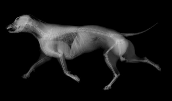
Radiographs (X-ray’s) and Ultrasound are valuable tools in veterinary medicine. Both are a painless, safe and non-invasive diagnostic tool that can help the doctor make a correct diagnosis for your pet.
Radiographs can be used to evaluate bones as well as the size, shape and position of many organs. Tumors can also, sometimes be detected by radiograph. It is used to diagnose bladder stones, broken bones, chronic arthritis, some spinal cord diseases and a variety of other conditions.
Ultrasound is different than radiation, in that, it uses high frequency sound waves and their echo to see an image. This enables us to see the structure as well as see inside of it. With Ultrasound we are better able to diagnosis various disesases including, heart disease, kidney and bladder disease, liver problems and diseases of other major organs. As an example, we can look inside the bladder for stones or growths. Internal abnormalities such as masses, cysts and abscesses can be seen, identified and measured.
Often radiography or ultrasound is used during your pet’s pregnancy to monitor the growth and number of puppies or kittens.
When needed, we send imaging to a specialist for further evaluation and consultation.
